Wound Analyzing
The W.H.A.T. analysis component is tightly integrated with the core W.H.A.T. platform to allow a comprehensive documentation covering all relevant areas of todays' wound management. The analysis tool itself is therefore started inside your web browser and is therefore completely platform independent allowing for a maximum of customer flexibility.
- Quality control for doctors and nurses in clinical and outpatient use (prevent forensic consequences)
- Detect co-morbidities (infection, vascular blocks, nutrition)
- Proved abridgement of lying duration, cost saving
- Reduction of wound infection
- Standardized documentation of doctors and nurses (input request for this document)
- Statistical evaluation and analysis (incidence of woundhealing disorders, duration of medical treatment)
- Planning requests for the use of bandaging materials and outlay (out-patient care, high-wastage)
- Objective evaluation of therapy (application observation, gauge for studies)
- Manifold uses in surgery, neurology, dermatology, geriatric care (decubitus, ischaemic laesion, diabetic foot, venous ulcus)
Process of the Wound Analysis
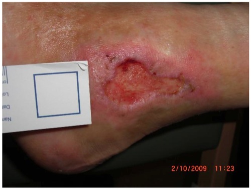
- Calibration
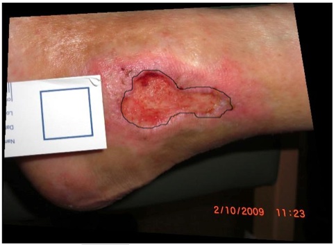
- Woundborder
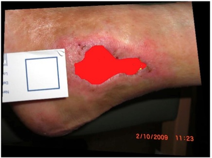
- Result
The following paragraphs will guide you through the process of performing a wound analysis inside the W.H.A.T. documentation platform.
- Step 1 Image Upload: To analyze an image, the user has to first upload it into the platform.
- Step 2 Image Dedistortion and Calibration: In order to be able to perform calculation on the image it has to be calibrated and dedistorted. For the user this process is kept as transparent as possible. All the user has to do is to indicated the location of the calibration square on the indicator stripe in the picture.
- Step 3 Wound Border Selection: The next step for the user is to indicate the wound size and location by tracing the edge/border of the wound with the mouse. Complex indications like selecting multiple wound areas or subtraction of two drawing areas to refine a specific area are also possible.
- Step 4 Wound Analysis: Using the given parameters W.H.A.T. performs the necessary calculations and provides the user both with a graphical result and a series of parameters (listed in detail in the next section).
- Step 5 Documentation: Both the graphical results and the numerical parameters are then saved into the W.H.A.T. documentation platform. From there it is possible to document further parameters (e.g. treatment strategies or used medical supplies) or to export the data either in a printable (e.g. PDF) or machine readable (e.g. CSV, Microsoft Excel) format.
Results
The following numerical parameters are automatically calculated from the wound image:
- Circumference (mm)
- Area (mm2)
- max. Length (mm)
- max. Height (mm)
- Granulation abs. (mm2)
- Fibrin abs. (mm2)
- Necrosis abs. (mm2)
- Granulation rel. (%)
- Fibrin rel. (%)
- Necrosis rel. (%)